
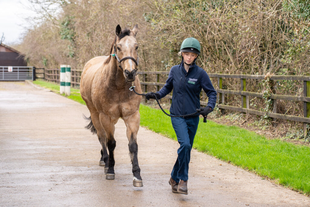
What is involved?
While in some cases the cause of lameness may be easy to elicit by clinical examination and gait evaluation, in others, more extensive investigations are required.
This may include diagnostic analgesia (“nerve blocks”), in which a local anaesthetic solution is injected around a nerve or directly into a joint or a synovial cavity, thereby temporarily desensitising a particular area of the limb.
Following the injection of local anaesthetic solution, the horse’s gait is reassessed to determine whether there has been improvement in the lameness.
This enables the veterinary surgeon to determine which part of the limb is painful and contributing to lameness.
In most lameness investigations, diagnostic imaging is used to help determine the cause of lameness.
The type of imaging used will depend on the nature of the lameness, but is likely to include radiography and ultrasonography.
In some cases, advanced imaging modalities such as MRI (magnetic resonance imaging) or nuclear scintigraphy (“bone scan”) may be indicated.
For these, we would refer you to another hospital which is equipped with these specific facilities.
Our lameness locator
In 2017, we invested in a new tool for lameness investigation at the clinic, the Equinosis Q inertial sensor lameness system (lameness locator).
The Equinosis Q system offers precise and objective identification of gait asymmetry using microelectric sensors positioned on the poll, pelvis and pastern of the horse.
The system is particularly useful in the investigation of subtle or multi-limb lameness, as well as to gauge the effectiveness of treatment.
It is also helpful when monitoring rehabilitation programmes.
The system is used in combination with standard lameness investigation techniques and can be used on all surfaces.
Treatment
Once the cause of lameness has been established, we will be able to advise you on appropriate treatment and management strategies.
This may include medication, surgery, farriery and exercise programmes.
Poor performance
Loss of performance in a competition horse can occur for many reasons.
The most common of these are musculoskeletal disease (lameness or back pain), upper respiratory tract disorders (“wind” problems) and lower respiratory tract disease.
Occasionally, internal medical problems (such as anaemia, viral infection, post-viral syndrome, gastric ulceration or cardiac disease), gynaecological problems (particularly in mares), dental issues or tack-related problems may be involved.
Investigation of poor performance can be challenging and involves multiple different diagnostic steps.
Advanced equipment, including telemetric ECG, gastroscopy and overground endoscopy, is available at the hospital.
We can take the overground scope and ECG equipment out to nearby gallops if we are investigating poor performance in racehorses.
Our on-site laboratory means that in-house analysis and same-day results are attainable for blood and other samples.
It is important that the horse’s normal tack is brought in and, where possible, that the horse’s usual rider is available at the time of initial consultation to ride the horse.